Clinical Kit 13/12/2012 Red Flag Tumour 2
The other case studies in this series can be access via;
Red Flag Tumor 2
23 February Physiotherapy
S/E October prior year developed L lumbar pain. No recalled trauma or reason for onset. In January started getting tingling sensation in L) lateral, anterior and posterior thigh, calf and foot. Also present in R but not as bad. L) foot not able to lift off ground. Now February has constant pain 3-4/10, increase with turning or sudden movements - can take breath away.
Has been seeing Chiro and OT for the past 2 months but has increased pain. Not pacing self or applying specific rest ratios. 36 yo female + 2 young children
Prior Lumbar XR taken, reported as normal by client (Doesn’t have or know where it is). No Ca PMH.
O/E NRPS 5/10
Active Movements: Flex to tibial tubercle, Ext 1/2 increase pain, SF L 5 above KJL increase pain, SF R 2 above KJL increase pain.
Palpation: L5 painful
Neurological: L34, L5S1 and S12 reflex present, power reduced L4 to S2 L), sensation decreased L) anterior, posterior and lateral thigh & lower leg.
Barbinsky and clonus normal. B & B function normal.
Nerve tissue provocation tests: mixed findings with SLR, cervical flexion test and slump test R & L.
Goal & Expectation: Find out what is happening and get relief
Treatment: Home Program/Advice Details:
- Explanation of need for diagnosis rather than just treat as before
- Need to commence some strength program, explain rest ratio, sleep posture and minimise loading
- Arrange XR (5 months prior, no report and situation since deteriorated)
26 February Physiotherapy
S/E Has had lumbar XR. No official report, but looks normal.
Sleeps good position. Rest ratio 30%. Lying down definitely reduces pain across L hip, but sensations same into both feet.
O/E TA all fours upper abdominal dominate pattern
Palp L5S1 > L45
Treatment: Bilateral dry needling for pain relief L45S1
Home Program/Advice Details: Commence gentle lumbopelvic strength exercises
Recommend seeing GP immediately and sent letter that day
4 March Physiotherapy
S/E Pain was less in lumbar spine for a couple of days but has built up again. P & N sensations stayed the same. Is traveling to Perth for family reasons. I suggested phoning GP to request MRI referral and have taken while in Perth on the weekend.
O/E Neurological: Power L weakest at foot eversion (L5S1), with weak tibialis anterior (L45), EHL (L5). Plantarflexion (S23) and knee extension (L34) reasonable. R eversion weak also but not as bad as L. Reflexes brisk L34, L5S1 and S12.
Treatment: Bilateral dry needling for pain relief L45S1
Home Program/Advice
- TA all fours + shoulder flexion with mirror, quite weak - practice
- TA + Clam shell for lumbopelvic strength and awareness
March MRI Findings
There is a large intradural extramedullary mass within the spinal canal at the T10/11 level. This lies dorsolateral to the cord on the left resulting in displacement of the cord anteriorly and to the right with marked associated cord flattening. There is no significant cord oedema or syrinx (intramedullar cyst). The mass is of intermediate signal intensity on T1 and T2 weighted images. It spans a craniocaudal distance of 18mm and measures up to 12.5mm in AP diameter. The lesion is most likely to represent a spinal meningioma.
At L5/S1, there is mild to moderate disc degeneration with mild posterior disc bulging. A shallow central 3 to 4mm posterior bulge/protrusion is present abutting the ventral theca. There is no evidence of nerve deviation or compression. The facet joints appear within normal limits.
Phone Follow-ups
- March underwent surgery for removal of benign spinal menigioma. Now has full strength but altered sensation - seems warm/hot in left leg. Leg is still quite achey when busy and during colder weatherJuly MRI follow-up - okayJan MRI follow-up - okay
What is a meningioma?
A meningioma is a tumor that develops from the meninges. Most meningiomas (90%) are categorized as benign tumors, with the remaining 10% being atypical or malignant.As benign tumors grow they can constrict and affect the brain/spinal cord tissue.
In many cases, benign meningiomas grow slowly, allowing neural tissue adaption. Depending upon where it is located, a meningioma may reach a relatively large size before it causes symptoms. Meningiomas growth rates are highly variable.
Most meningiomas are singles, but it is possible to have simultaneous tumors in different parts of the brain and spinal cord.
What are spinal meningiomas
Meningiomas account for approximately 25% of all spinal tumors. Most spinal meningiomas are intradural (within or enclosed by the dura mater) and extramedullary (70% outside or unrelated to medulla). They can cause a mix of back pain, radiculopathy and myelopathy symptoms, producing a seemingly confused clinical picture.
Meningiomas are the second most common tumor in the intradural, extramedullary location, second to tumors of the nerve sheath.
Approximately 80% of spinal meningiomas are located in the thoracic spine, followed by cervical spine (15%), lumbar spine (3%), and the foramen magnum (2%).
Who is at risk?
Meningiomas are most common in people between the ages of 40 and 70. They are more common in women than in men. Among middle-aged patients, there is a marked female bias, with a female: male ratio of almost 3:1 in the brain and up to 6:1 in the spinal cord.
Common Examination Findings
On physical examination, sensory and motor deficits are seen almost equally. A high incidence of Brown-Sequard syndrome is seen (ipsilateral paralysis, decreased tactile and deep sensation, and a contralateral deficit in pain and temperature sensation). This finding is most likely secondary to the high incidence of laterally positioned meningiomas.
With substantial growth of the tumors, clinical findings may merge. Signs and symptoms often are sequential, with weakness and stiffness of the legs typically preceding pain and the occasional findings of bowel and bladder changes.
Treatment
The duration of symptoms may span 6-23 months. Failure to accurately and efficiently identify patients with myelopathy can result in progression of symptoms that are no longer effectively treated with conservative or surgical interventions.
Resection of spinal meningiomas can result in excellent recovery, even in patients with notable preoperative deficits. The surgical morbidity rate is low because surgical resection of a meningioma can easily be accomplished via laminectomy.
What did I Learn?
Essentially, red flags are signs and symptoms found in the patient history and clinical examination that may tie a disorder to a serious pathology. As primary contact practitioners we need to screen for and act upon S & S that indicate possible red flag pathology. The constant pain, worsening symptoms, variable bilateral parathesia, non responsive nature to past treatment and foot drop from the history, all point towards possible significant pathology, but is it a red flag?
The more common possibility would be a moderate L) postrolateral to central IVD protrusion. This could cause the lower motor neuron (LMN) symptoms of lumbar pain, radiculopathy L > R, increased sensitivity of neural tissue provocation tests, reduced motor testing and function and normal B & B function. It could also cause the lack of remission and lack of success from 2 months manipulative treatment (chiropractic) due to the chemical irritation and lack of pacing for a significant IVD protrusion (although the time frame was too long for my clinical comfort).
However as noted in my first treatment, there was no clinical diagnosis.
I felt I needed to consider a tumor. On the -ve, the client was presenting with unremitting pain (however with a mechanical element) that was non responsive to conservative treatment. On the +ve she was not over 55 years, there was no unexplained weight loss, and no past history of cancer. However there was also the range of mixed neurological signs and symptoms.
When differentiating between an upper (cord compression myelopathy) and lower (radiculopathy) motor neuron pathology, observance of superficial and deep reflexes is beneficial. The deep tendon reflexes of patella, medial hamstring and achilles were brisk bilaterally, but Barbinski and clonus were -ve. Being suspicious of an UMN lesion, on review, I could also have checked the superficial reflexes;
- Abdominal - Epigastric, Upper Abdominal and Lower Abdominal (T8-12)
- Cremasteric (L1-2) - if a male client
because a person with an UMN lesion (compression of corticospinal tract) will generally present with absent superficial reflexes and hyper reflexia of the deep tendon reflexes. I could have also examined contra lateral pain & temperature deficits checking for a Brown-Sequard syndrome.
Another feature of an UMN lesion is the non-dermatonal distribution of sensory changes compared to a LMN lesion; as was present in this case. Especially in someone so young, you wouldn't expect spinal degenerative changes to cause myelopathy signs and symptoms.
In Summary
For me the ongoing symptoms (time and treatment factors) and mixed neurological findings (foot drop, brisk reflexes and multi-dermatonal sensory presentation) was enough to raise my clinical suspicions and arrange an XR and then MRI as quickly as possible. Being in a rural and remote practice, we have to take opportunities as they present, like social trips to Perth.
An interesting hypothetical to consider; if only a lumbosacral CTS or MRI had been arranged and the 3 to 4mm posterior bulge abutting the ventral theca at L5/S1 had been identified, would that have satisfied you clinically?
As a general note when performing NTPT, apply only light longitudinal load for very short periods of time to avoid neural elongation and consequential compression of intraneural vascular tissue and subsequent ischemia.
Recent Blogs of Interest
- Frustrated with Home Exercise Program Compliance? - VideoXs Solution
- Updated list of courses in Perth and Brisbane for 2013 includes Student Pizza & PD nights, Anatomy Workshops, Mulligans, RT & MSK US, Complex Necks, Podiatry and OT Intensive programs
- Red Flag Tumor 1
All the best,
Doug Cary FACP
Specialist Musculoskeletal Physiotherapist
email doug@aapeducation.com.au
ph/fx 08 90715055
Receive a FREE Information Report
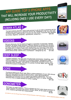
Choose The Top 5 Manual Therapy Apps or Infection Control & Needling (V2)
Along with the report you'll also get a complimentary subscription to "Clinical Kit," our regular eZine (email newsletter) and Free Bronze Membership. You'll get ideas, information, insight and inspiration on a regular basis, plus access to our Resource Library, helping you unravel those clinical conundrums appearing every day.
You are free to use material from the Blog in whole or in part, as long as you include complete attribution, including live website link. Please also notify me where the material will appear. The attribution should read: "By Doug Cary FACP of AAP Education. Please visit our website at www.aapeducation.com.au for additional clinical articles and resources on post graduate education for health professionals" (Please make sure the link is live if placed in an eZine or in a web site.)
HOME | DRY NEEDLING | ANATOMY WET LAB |INTEGRATED NECK | 100% GUARANTEE | OUTCOME MEASURES | MSK & RTUS | BLOG

